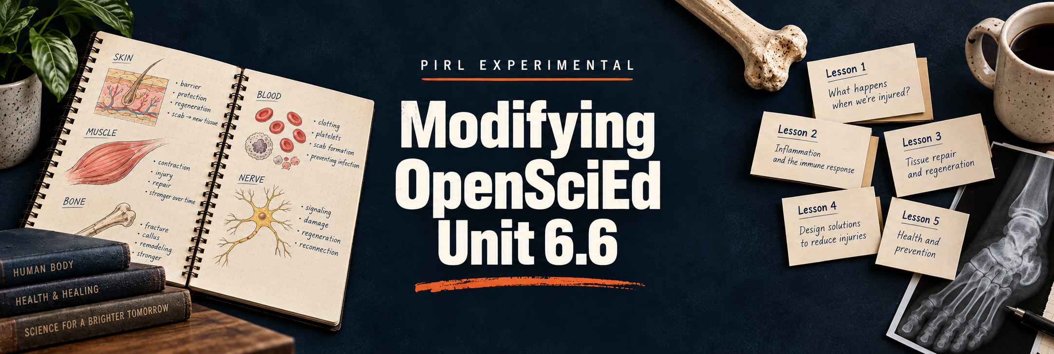
PIRL Experimental · Modifying OpenSciEd Unit 6.6
Explore the series
Classroom decisions, practical modifications, and work still in progress. Select a title to explore the chapter, then follow its link to read the full page.
Overview The Curriculum Is Not the Teacher
A published curriculum can offer a strong storyline, useful evidence, and ambitious goals while still needing substantial changes for the students in front of us. This introduction explains the thinking behind my modifications to OpenSciEd Unit 6.6: Injuries & Healing. I look at what deserves protection—students asking questions, interpreting evidence, building models, and connecting ideas—and what can change when a procedure, discussion, or written task takes more time than the learning justifies. The shift to 66-minute periods makes those decisions especially visible, but the underlying question reaches beyond any particular schedule: What do students need to understand, and what will help them get there?
The overview also establishes the tensions that run through the series: inquiry and direct instruction, pacing and coherence, accessibility and intellectual demand, and curriculum fidelity and teacher judgment. These chapters document classroom experience, unresolved problems, and possible next steps. They explain the reasoning behind each modification so another teacher can decide what fits their own classroom. For the principles guiding the lesson-by-lesson work, begin with The Curriculum Is Not the Teacher.
Lesson 1 Start With the Mystery
The opening lesson needs to make an injury interesting before students know enough anatomy to explain it. In this chapter, I rethink OpenSciEd's four-day anchoring sequence for a schedule that gives me, at most, two 66-minute periods. Photos, X-rays, recovery evidence, and visible changes in function give students something concrete to notice before they encounter dense medical reports. Guided reading then helps them confirm or revise their first ideas. The goal is to preserve the mystery of M'Kenna's injured foot while reducing the procedures and reading demands that can obscure the important clues.
I examine how a supplied healing timeline, layered foot diagram, evidence-and-reasoning card sort, and more efficient collection of student questions can support that opening storyline. A short diagnostic can reveal what students already think without requiring a lengthy assessment. Throughout the redesign, I want students to connect damaged structures, changes over time, and returning function while recognizing that they still cannot explain how healing happens. Lesson 1: Start With the Mystery follows the choices behind that launch and offers condensed and practical guides for planning it.
Lesson 2 From a Wing to a Working System
Students need to see a limb as an interacting system before they can explain why an injury changes what it can do. This chapter describes why I replaced the chicken-wing comparison with a frog hind leg, whose lower leg and foot offer a more immediate connection to the human injury anchoring the unit. A guided investigation makes the relationship between muscle contraction, bones, and movement visible. Repeating a pull after damaging a bone helps students examine how a change in one structure affects the function of the larger system, rather than simply naming parts they found during a dissection.
The discussion also considers mechanical models, splints, and stabilization as possible extensions of that reasoning. Those experiments raise a useful planning question: Does an added activity clarify how the system works, or simply give students more to do? I separate the established frog investigation from models I am still developing, account for handling and cleanup time, and connect the work back to the layered foot organizer. Lesson 2: From a Wing to a Working System explains how I am protecting the structure-and-function story while deciding which models earn a place in the lesson.
Lesson 3 Learning to See Inside the Body
An X-ray, an MRI, a cross section, and a textbook diagram may show related structures, but students do not automatically know how to connect them. This chapter examines why a seemingly short medical-imaging lesson routinely expands to fill much more class time. The challenge is learning to interpret unfamiliar representations while also reading, observing, drawing, and recording ideas. I reconsider how much students should produce from every image and how explicitly I need to teach the moves that help them connect a real medical image with a more accessible diagram.
The protected reasoning chain leads students from images of body structures to the recognition that blood vessels and nerves extend through skin, muscle, and bone. I explore a more focused observation routine, purposeful medical-imaging reading, and scaffolded labeling when drawing would add work without adding understanding. I also consider whether a shorter version could move earlier in the sequence to establish useful anatomy before the dissection. Lesson 3: Learning to See Inside the Body traces those decisions while leaving the functions of blood and nerves available for students to investigate in the lessons that follow.
Lesson 4 Blood — The Lesson I Haven't Solved Yet
This chapter begins with a dependable lesson and an unresolved question: Could a more active model make blood's composition and functions easier for students to understand? The existing reading provides a useful baseline for teaching red blood cells, white blood cells, platelets, and plasma, along with their roles in transport, injury, and healing. A reciprocal-teaching routine asks students to predict, question, clarify, and summarize. Moving microscope skills into separate instruction also helps keep the blood lesson focused on its own scientific purpose instead of trying to teach tool use and content at the same time.
From there, I consider several possibilities, including a Bag of Blood model, component stations, a circulation role-play, a commercial kit, and a larger skeleton-based model. These are candidates to pilot and evaluate, not a finished replacement sequence. The chapter asks what each approach makes visible, how much time it requires, and whether it helps students connect a component's structure with its function. Lesson 4: Blood — The Lesson I Haven't Solved Yet offers an honest account of lesson development when the current approach works, but a more useful one may still be possible.
Lesson 5 Signals in Motion
A second reading-centered lesson can feel very different when it immediately follows a reading about blood. This chapter considers how to make nervous-system signaling something students can physically trace, test, and explain. A human relay models a message moving from a receptor through a pathway to a brain group, then outward toward a response. Blood-review questions can become part of that relay, connecting retrieval of earlier learning with the new model. Selective direct instruction helps students distinguish the receptor, sensory pathway, brain or spinal cord, motor pathway, and responding muscle without letting the activity become a loose game of telephone.
Reaction-time comparisons, a ball-catching example, and questions about damaged pathways bring the model back to sensation, response, numbness, and the delayed return of feeling in the injured foot. I also discuss demonstrations I am considering and why some belong in a smaller pilot before becoming part of the default lesson. The choice between a physical model and a reading remains connected to what happens in Lesson 4. Lesson 5: Signals in Motion follows that planning process and keeps the focus on signals moving through a connected network.
Lesson 6 From Messy Evidence to Useful Models
Students can spend a productive-looking period at microscopes and still leave with drawings that reveal little about what they noticed. This chapter reconsiders what students should do with microscopic evidence from skin, muscle, bone, blood, nerves, and adipose tissue. Separating basic microscope instruction from the tissue investigation creates room to focus on the evidence itself: boundaries, repeated patterns, differences among tissues, and the relationship between a real view and an idealized diagram. The central question is whether a student's recording helps them build an explanation or simply rewards their ability to produce a recognizable picture.
I explore alternatives to drawing every specimen, including mystery-sample matching, annotated observations, comparisons across representations, and a scale sequence from whole body to tissue to cell. I also consider where the investigation might fit most usefully in the unit, while being clear that those changes are still under development. Students should leave with a simple, usable model that connects injured body structures to a cellular explanation of healing. Lesson 6: From Messy Evidence to Useful Models examines how to protect authentic observation while making the thinking, rather than the drawing task, the main product.
Lesson 7 Assessing the Story We Actually Built
At the midpoint of the unit, I want to know whether students can explain the injured foot using the ideas we have actually developed together. This chapter describes why I departed from OpenSciEd's midpoint task and built an assessment around skin, muscle, bone, blood, nerves, and their interactions. The assessment asks students to apply structure-and-function reasoning to the anchoring phenomenon, helping me distinguish connections they can use independently from ideas that only sounded secure during class discussion. That makes the checkpoint useful for deciding what to revisit before the unit moves more deeply into cellular healing.
One open-response assessment gradually became a family of versions with different levels of support: visible success criteria, more focused prompts, sentence frames, reduced writing demands, selected responses, and digital formats. I examine what those changes reveal and what they may change about the evidence an answer provides. Recognizing a correct explanation is different from generating one, and difficulty organizing a written response is not automatically a gap in science understanding. Lesson 7: Assessing the Story We Actually Built explains the assessment choices and how I weigh support against the thinking I need students to demonstrate.
Lesson 8 From the Gap to the Mechanism
Students have seen that an injury can heal, but an improved appearance does not explain what happened in between. This chapter protects a strong OpenSciEd move: using a torn tissue model to make the missing material visible and turning that gap into a question about cells. A paper model taped into the notebook creates flaps students can open and examine. Time-lapse evidence then gives them changes to explain, moving the discussion from the visible wound toward possible mechanisms operating at a smaller scale. The lesson should make a cellular explanation necessary without supplying the formal answer too soon.
My proposed refinements focus on how much thinking students do before scaffolds or candidate explanations are introduced. An independent prediction can reveal more than choosing from options immediately, while a purposeful comparison of ideas can prepare students to revise their models. I consider shortening recording sheets, question-board routines, and extended sharing while preserving the injury gap, the evidence, and the unresolved mechanism. Lesson 8: From the Gap to the Mechanism shows how I am trying to create a clear need for the cell-division investigation that follows without overbuilding a lesson whose central idea already works.
Lesson 9 From Watching Cells Split to Explaining Healing
After students predict how a gap in damaged tissue might be filled, they need evidence for a mechanism that can produce additional cells. This chapter examines the use of cell-division videos and images to connect observation with an explanation of healing. The instructional focus is on a usable sequence: a cell grows and copies its internal material, that material separates as the cell begins to divide, and two cells result. Formal phase names are not the destination. Students need to use what they observe to explain how existing cells can give rise to more cells in damaged tissue.
The chapter also questions how much of that explanation I have been doing for students. A growing collection of mini-lessons, labeled diagrams, fill-in-the-blank organizers, and answer keys can make the path clear while leaving too little for students to construct. I consider a sequence that moves from video evidence to a card sort, a shared model, and an independent labeled explanation. Lesson 9: From Watching Cells Split to Explaining Healing follows that reassessment of scaffolding and keeps the final task connected to the original injury: explaining how an increase in cells could help close the gap.
Lesson 10 From Cell Lifespans to Explaining Whole-Body Healing
A short reading about cell lifespans became the center of my version of Lesson 10, but later assessment showed that students were not always extending the healing explanation beyond skin. This chapter examines how to make that transfer more explicit. An infographic provides patterns to investigate: different cells persist for different lengths of time, and tissues can maintain themselves while individual cells are replaced. Students then return to the tissues involved in the injured foot and consider how cell production relates to repair and growth throughout the body, rather than treating cell division as a skin-only explanation.
I describe what I cut from the longer lesson and what reasoning the shorter version still needs to preserve. Cell-lifespan data offer clues, but they do not provide a simple ranking of healing speed, and students should not infer that every mature specialized cell routinely divides. The work is to make a supported prediction, identify what the available evidence can explain, and recognize what else needs investigation. Lesson 10: From Cell Lifespans to Explaining Whole-Body Healing develops that bridge from a memorable data display to a more connected explanation of tissue maintenance, injury, and growth.
Lesson 11 From Cell Membranes to Cell Delivery
This is a lesson I have often cut, and this chapter asks what it would take to give it a protected place in the unit. The central investigation uses red onion cells observed in plain water, salt water, and plain water again to examine changes associated with water moving across cell boundaries. A procedure video and reference images can support or rescue the investigation when microscope work becomes difficult. The purpose is to help students reason about a cell's boundary and the movement of materials it needs, while keeping that work connected to the larger injury-and-healing story.
I then consider a delivery model that connects blood vessels and capillaries with nearby tissue cells. That change in scale can help students explain how materials traveling through the body become available where repair is happening. A simulation or reading may support the explanation, but each addition needs a clear role. The chapter also addresses assessment fairness: detailed membrane content should not remain on a test if the related instruction was omitted. Lesson 11: From Cell Membranes to Cell Delivery sets out the investigation, possible supports, and the connection I want students to make between circulation and cellular needs.
Lesson 12 From Human Healing to Salamander Transfer
An unfamiliar organism can make students feel that an assessment is asking about something they were never taught. This chapter describes how a salamander regenerating an arm became an opportunity to practice using familiar science in a new context. Its limb offers a useful comparison with the human injury: students can consider skin, muscle, bone, blood, nerves, and cells while recognizing that the outcome is different. Repeated video observations before an explanation give them evidence to inspect, and models at the body-system and cellular scales ask them to connect that evidence with ideas developed across the unit.
I treat the task as a practice transfer assessment, protecting an independent attempt before feedback, examples, and revision. The goal is to help students identify what they already know, decide how it might apply, and name questions that remain. They are not expected to supply advanced regeneration mechanisms the unit has not taught. I also examine how to preserve that thinking in a shorter period by reducing extended discussion. Lesson 12: From Human Healing to Salamander Transfer explains the classroom sequence, the limits of the comparison, and possible next steps for a more deliberate transfer task.
Lesson 13 Assessment Modification Without Changing the Target
Changing an assessment format can remove a barrier, change the science being assessed, or do both. This chapter examines that distinction through the unit's summative transfer work on growth. Students bring ideas about cells, tissues, cell production, and material delivery to evidence involving growing bodies, growth plates, and plants. The challenge for the teacher is to identify which scientific connections students must demonstrate and then decide whether the original reading, writing, organization, and response demands help reveal those connections or make them harder to see.
I compare a family of assessment versions, including open responses, scaffolded organizers, sentence frames, word banks, partially completed responses, and digital forms. Rubrics and visible success criteria help clarify the destination, but supports still need scrutiny: selecting an explanation provides different evidence from constructing one, and a prompt can sometimes supply the reasoning it was meant to assess. The chapter offers a practical process for choosing support and being explicit about any change to the target. Lesson 13: Assessment Modification Without Changing the Target explores how to make an assessment more accessible while remaining honest about what the resulting answers show.
Lesson 14 Saving the Lesson That Always Gets Cut
The final lesson asks students to extend the unit beyond a biological account of injury and healing, yet it is also the lesson I have repeatedly run out of time to teach. This chapter begins with that admission and considers how to preserve its purpose within a realistic schedule. Returning function, a changed body, and a person's experience are not interchangeable ideas. First-person accounts of disability can help students attend to agency, visible and invisible differences, and the ways environments enable or restrict participation. Those perspectives need a meaningful place in the unit's conclusion.
I outline possible versions ranging from a full 66-minute arc to a shorter sequence centered on one person's story, one access barrier, and one proposed redesign. An accessibility audit, photo walk, or focused design task could help students move from listening to examining their surroundings, provided they do not substitute assumptions for the perspective they just encountered. These are designs to try, not claims about a finished lesson. Lesson 14: Saving the Lesson That Always Gets Cut considers what to protect, what to shorten, and how the unit might end with students thinking carefully about function, access, and design.