Pirl Experimental · Interactive Science
Interactive Science
Simulations
Explore blood, cells, body systems, healing, photosynthesis, the microscopic world, and coastal flooding. Find a model, test an idea, and use your observations to build an explanation.
14 of 14 simulations
Open a simulation or expand its classroom ideas.
Coastal Science & Engineering
Coastal Flood Lab
Can you keep the water out? Test coastal defenses against waves, high water, and rain. Explore Point Pleasant Beach and Borough, compare designs, and use modeled flood depth and water volume as evidence.
- Demo · Standard · Honors
- Two local towns
- Test coastal defenses
- Export trial data
Start with the Demo wave tank, then investigate neighborhood flooding in Standard or Honors. This educational model does not predict flooding at specific properties.
Open simulationClassroom ideas
- Run a fair test: In Demo, compare two defenses with the same ten waves. Count how many waves carry water over each barrier.
- Compare the towns: In Standard, test the same storm in the Beach and Borough cross sections. Explain how each model's landscape changes the path of water.
- Design within a budget: In Honors, choose defenses and drainage improvements, then evaluate their effect on road depth and neighborhood flooding.
- Use the evidence: Export Standard or Honors trial data, compare results with a prediction, and explain one limitation of the model.
 Body ExplorerOpen the interactive explorer.
Body Systems, Tissues & Cells
Body Explorer
Explore skin, nerves, blood, fat, muscle, and bone. Move among naked-eye, tissue-diagram, microscope, and cell views to compare what becomes visible at each scale.
- 6 body structures
- 4 levels of view
- Zoom slider
- Compare & sketch
Connect tissue structure to injuries and healing. Use the models alongside classroom microscope slides to compare what you can see.
Open explorerClassroom ideas
- Choose a tissue and sketch the patterns you notice in its diagram and microscope views.
- Compare two tissues using the same features: cell shape, arrangement, and the spaces or material between cells.
- Sketch what a cut, tear, or fracture could change in the tissue. Label the evidence behind your choices.
- Explain what would need to be repaired for the tissue to do its job again.
 Microscope ExplorerOpen the interactive explorer.
Microscopy & Observation
Microscope Explorer
Look closer at 12 materials and prepared specimens, including crystals, a printed letter e, feathers, cotton fibers, hair, a flea, and a fruit fly. Compare naked-eye and microscopic views with a zoom slider.
- 12 samples
- 2 levels of view
- Zoom slider
- Observation practice
Illustrated teaching models support real microscope work. Compare materials that were never alive with samples that came from living things.
Open explorerClassroom ideas
- Predict which details will become visible before moving the zoom slider.
- Sketch the same sample in both views. Label details you could only see after zooming in.
- Compare two samples using shape, texture, and repeating patterns as your evidence.
- Use the printed letter e to discuss image orientation, then compare its view with a real compound microscope.
 Muscles Work in PairsExplore the arm and foot simulator.
Body Systems & Movement
Muscles Work in Pairs
Explore how opposing muscle pairs move the arm and foot. Switch between elbow and ankle models, choose a movement, and watch one muscle shorten as its partner lengthens.
- Arm & foot models
- Antagonistic pairs
- Contraction & relaxation
- Play & pause
A simple animation for exploring how muscles, tendons, bones, and joints work together. Muscles pull; they do not push bones.
Open simulatorClassroom ideas
- Predict which muscle will shorten before choosing a movement.
- Pause the cycle and compare the shapes of the contracting and relaxing muscles.
- Compare the elbow and ankle models and identify the pattern they share.
- Use observations to explain why an opposing muscle is needed to move a joint in the other direction.
 Blood: A Working SystemOpen the interactive virtual lab.
Cells, Body Systems & Injuries
Blood: A Working System
Investigate blood from the microscope slide to injured tissue. Prepare a virtual smear, separate blood's components, trace materials through capillaries, and model clotting, swelling, immune cleanup, and tissue exchange.
- Virtual blood smear
- Mixture separation
- Capillary exchange
- Clotting response
Built for OpenSciEd Unit 6.6 Lesson 4, with evidence-log prompts that connect blood's components to transport and injury response.
Open virtual labClassroom ideas
- Compare mammal and nonmammal blood cells under the virtual microscope.
- Separate the mixture and explain why whole blood can look uniform even though it contains different components.
- Trace oxygen, nutrients, water, electrolytes, carbon dioxide, and wastes across capillary walls.
- Build an evidence-based sequence showing how plasma, platelets, white blood cells, and continued blood flow respond after injury.
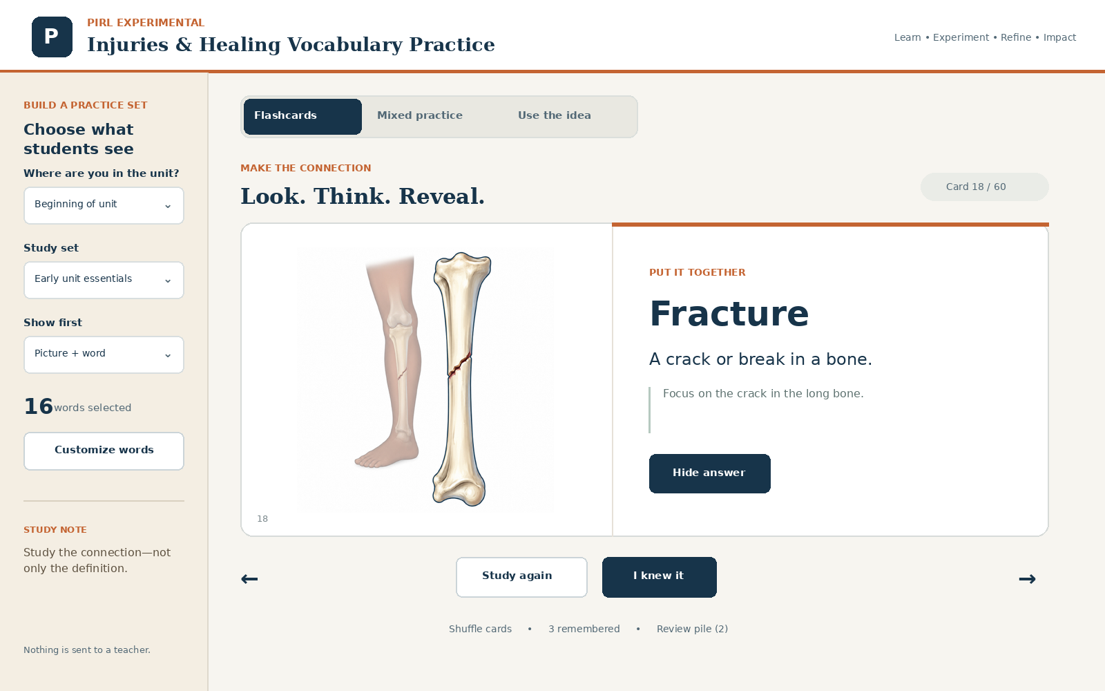 Injuries & Healing Vocabulary PracticeOpen the interactive practice tool.
Cells, Body Systems, Injuries & Imaging
Injuries & Healing Vocabulary Practice
Practice the unit vocabulary through illustrated flash cards, mixed retrieval, and short application situations. Filter by lesson stage, choose what appears first, shuffle the set, and revisit words that need more work.
- Illustrated flash cards
- Mixed retrieval
- Application practice
- Review pile
Uses the same teacher-reviewed terms, definitions, and images as the printable Injuries & Healing word wall.
Open practiceClassroom ideas
- Filter to the current lesson so students practice only the words they have encountered.
- Use picture-first and definition-first rounds to vary the direction of retrieval.
- Assign the review pile for a short warm-up, station, or targeted support session.
- Pair the digital practice with the matching wall cards or notebook flash cards.

Body Systems, Injuries & Imaging
Mystery Injury Simulator
Examine the injured foot and illustrated X-rays, then change the object, mass, and drop height to investigate how the injury may have happened. Explore injury and recovery cards, and keep evidence alongside your thinking.
- Examine the foot
- Recreate the injury
- 27 reference cards
- Copyable evidence log
A simplified teaching model for an Injuries & Healing unit launch. Similar evidence can support a possible explanation without proving the exact drop.
Open simulationClassroom ideas
- Examine the original case. Record the type and location of each piece of evidence, along with initial thinking or questions.
- Change one drop variable at a time, then use the same examination tools to compare each trial with the original injury.
- Explore fracture types, bruising, swelling, scars, and recovery devices. Turn a card over, then save a useful reference with a question or explanation.
- Copy the evidence log into a Form or Doc and use it to support an explanation, including what the evidence cannot establish.
Cells & Energy
Cell Membrane Transport Virtual Lab
Explore simple diffusion, facilitated diffusion, and active transport. Change molecule counts, control selective channels, and use an ATP pump to investigate movement into and out of a cell.
- 3 transport pathways
- Visual hover previews
- Live particle counts
A simplified teaching model with stated assumptions. Use the starting examples to make predictions, compare movement in both directions, and explain the role of channels and ATP.
Open simulationClassroom ideas
- Start with equal counts and observe whether molecules continue crossing the cell membrane in both directions.
- Compare a blocked channel with an available channel. Explain why a difference in concentration is not always enough for transport.
- Use the ATP pump to move sodium toward the side that already has more sodium. Track how the counts and ATP supply change.
Plants & Photosynthesis
Leaf Disk Photosynthesis
Compare a reference cup and two treatments, watch disk infiltration, and analyze simulated flotation. Explore how a model connects photosynthesis, gas accumulation, and buoyancy.
- 3 comparison cups
- Adjustable variables
- High school biology
Theoretical model with explicit assumptions. Use alongside a real leaf disk assay; its simulated times are not expected classroom results.
Open simulationClassroom ideas
- Before a real assay, predict how changing one variable could affect flotation while keeping other conditions constant.
- Practice graphing simulated counts and interpreting ET₅₀, the estimated time for half the disks to float.
- Compare the model with actual class observations or a makeup investigation. Identify assumptions that may explain differences.
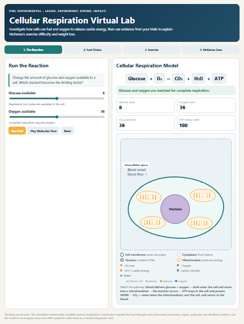 Cellular Respiration Virtual LabOpen the simulation to explore the model.
Cells & Energy
Cellular Respiration Virtual Lab
Follow glucose and oxygen from the bloodstream into a cell. Trace how the model represents ATP production and the movement of carbon dioxide and water.
- Cell processes
- Matter & energy
- Bloodstream connection
Classroom ideas
- Identify the materials entering and leaving the cell in the model.
- Explain how the bloodstream and cells work together.
- Distinguish the movement of matter from the model’s representation of energy transfer.
 Capillary & Tissue ExchangeOpen the simulation to explore the model.
Cells & Body Systems
Capillary & Tissue Exchange
Follow blood through a capillary bed as oxygen and nutrients move toward tissue cells and carbon dioxide and other wastes return to the bloodstream.
- Capillary bed
- Tissue exchange
- Pause & observe
Classroom ideas
- Pause the animation and describe where different materials are moving.
- Follow the same red blood cells through the capillary bed.
- Connect the exchange shown here with the needs of cells in the respiration model.
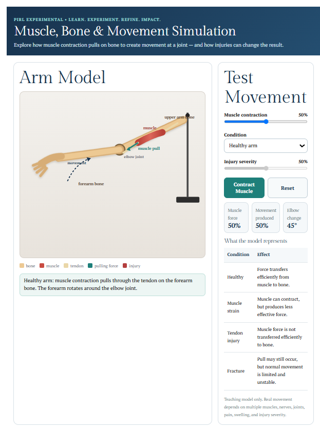 Muscle, Bone & MovementOpen the simulation to explore the model.
Body Systems & Injuries
Muscle, Bone & Movement
Contract a muscle, follow the force through the arm, and compare how muscle, tendon, and bone injuries change the movement shown in the model.
- Interactive arm
- Muscles & tendons
- Injury comparisons
Classroom ideas
- Observe the uninjured model and identify the structures involved in movement.
- Predict what will change when a muscle, tendon, or bone is injured.
- Use observations from the model to explain how connected structures work as a system.
 How Does a Cut Heal?Open the simulation to explore the model.
Cells, Injuries & Healing
How Does a Cut Heal?
Follow a cut from bleeding and clot formation to new cells filling the gap. Change conditions such as oxygen, nutrients, hydration, or infection and compare the modeled repair.
- Skin repair
- Cell division
- Compare conditions
Classroom ideas
- Describe what changes at the wound as the model progresses.
- Compare one changed condition with the model’s starting conditions.
- Use the cell-division close-up to connect changes in cells with repair of tissue.
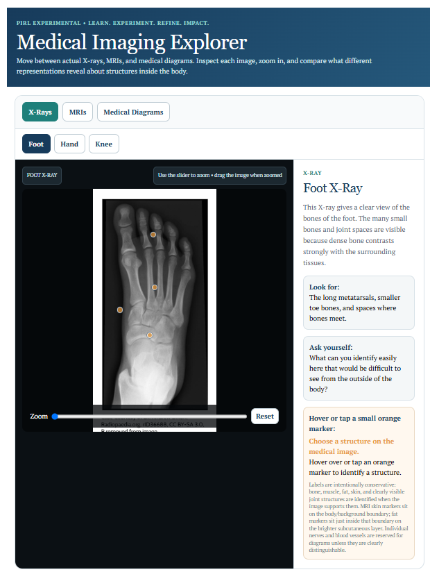 Medical Imaging InteractiveOpen the simulation to explore the model.
Body Systems & Imaging
Medical Imaging Interactive
Compare X-rays, MRI images, diagrams, and cross-sections. Investigate what different views reveal about structures inside the body.
- X-ray & MRI
- Cross-sections
- Visual comparison
Classroom ideas
- Compare two views of the same body region and identify what each makes easier to see.
- Connect a cross-section with the larger structure it represents.
- Separate what an image shows directly from an inference you make about it.
No simulations match this search and topic. Try a broader term or clear the filters.